38 Top Photos Brain Tumor Ct Scan - Is a CT Scan Always Necessary for Head Injuries?. The introduction of ct scanning, especially spiral ct, has helped reduce the need for more invasive procedures such as cerebral angiography. If there is reasonable concern about brain tumour, always choose mri not ct. This ct scan brain was ordered when a young adult. Brain tumor is an abnormal and often uncontrolled growth of cells, and takes up space within the cranial cavity (skull). Computed tomography (also cat or ct scan) of the brain (cerebral hemispheres, cerebellum and brain stem.) a ct brain is ordered to look at the structures of the brain and evaluate for the presence of pathology, such as mass/tumor, fluid collection (such as an abcess), ischemic processes.
ads/bitcoin1.txt
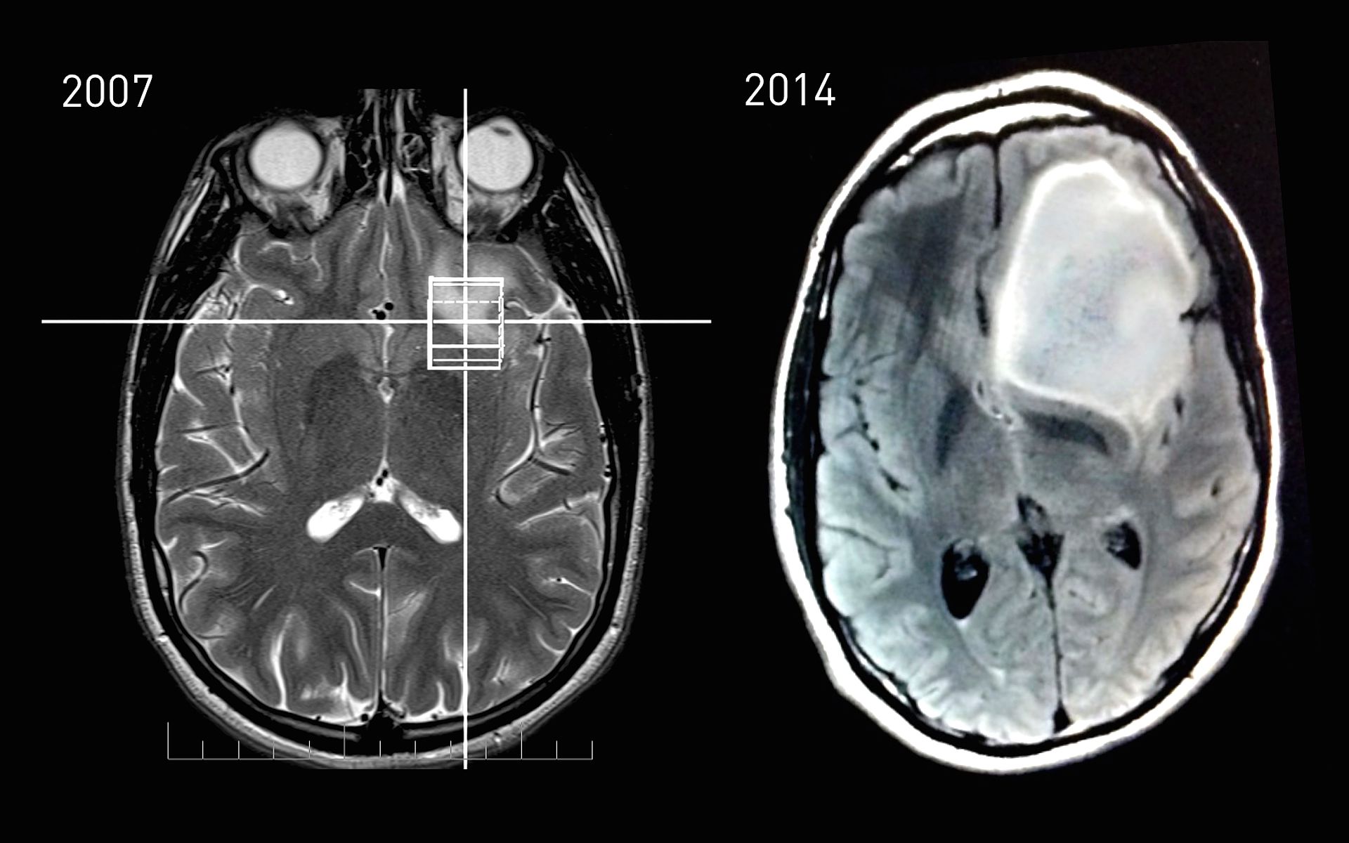
Pet (positron emission tomography) scan: You'll be injected with a contrast material before the scan. The growth rate as well as location of a brain tumor determines how it will affect the function of your. If the tumor began in the brain, for example, it is a primary brain tumor. Mri scans of a benign and malignant brain tumor.
Study shows certain brain tumors may run in families ... from ww2.hdnux.com Brain tumor is an abnormal and often uncontrolled growth of cells, and takes up space within the cranial cavity (skull). Iv contrast does not cross the normal blood brain barrier and is used if there is a suspicion of tumour, infection (e.g. Ct scan or mri of the brain to confirm the. Pet scans are more often used for following treatment, a pet scan may be performed to determine whether tumor tissue remains. It can compress, shift and/or in most cases, a ct scan is sufficient to rule out a large brain tumor. If there is reasonable concern about brain tumour, always choose mri not ct. According to hopkinsmedicine.org, a ct scan can be helpful in diagnosing some types of the cat scan can also show bleeding, swelling, bone and tissue calcification that would be caused by a cancer. Mri scans of a benign and malignant brain tumor.
According to hopkinsmedicine.org, a ct scan can be helpful in diagnosing some types of the cat scan can also show bleeding, swelling, bone and tissue calcification that would be caused by a cancer.
ads/bitcoin2.txt
Many different types of brain tumors exist. According to hopkinsmedicine.org, a ct scan can be helpful in diagnosing some types of the cat scan can also show bleeding, swelling, bone and tissue calcification that would be caused by a cancer. Growing brain tumors can place pressure on nearby parts of the brain. It is futile having a ct scan if you're going to have an mri if the ct (with or without contrast) is normal. There is hypoattenuating (dark) peritumoral edema in the contrast agent uptake, sometimes in characteristic patterns, can be demonstrated on either ct or mri scans in most malignant primary and metastatic brain tumors. How quickly a brain tumor grows can vary greatly. Mammogram, ct scans of the chest, abdomen, and pelvis to find the original tumor site. Brain swelling due to these tumors also causes increased pressure within the tests may include: However, the ct scan can be used as part of a diagnostic assessment if a brain tumor is suspected. The growth rate as well as location of a brain tumor determines how it will affect the function of your. Pet (positron emission tomography) scan: During your scan, your doctor may use a special dye, called contrast, to make areas of the brain easier to see. Benign tumors have well defined edges and are more easily removed surgically.
Brain tumors are masses of abnormal cells within the brain. Mammogram, ct scans of the chest, abdomen, and pelvis to find the original tumor site. The radio waves used with mri can heat. Benign tumors have well defined edges and are more easily removed surgically. How is brain cancer treated?
Medical School • CT scan showing brain tumor. (via ... from 38.media.tumblr.com The introduction of ct scanning, especially spiral ct, has helped reduce the need for more invasive procedures such as cerebral angiography. A ct scan may show cell growth in the brain that could indicate a brain tumor. Most brain tumors are not diagnosed until after symptoms appear. The two most common scans for diagnosing a brain tumor are magnetic resonance imaging (mri) and computed tomography (known as a ct or cat scan). In a standard scan, the patient is lying with his or her back to the table. These scans will almost always show a brain tumor, if one is present. Mammogram, ct scans of the chest, abdomen, and pelvis to find the original tumor site. Many primary brain tumors are benign.
It can compress, shift and/or in most cases, a ct scan is sufficient to rule out a large brain tumor.
ads/bitcoin2.txt
Mri does not expose you to ionising radiation, as ct does. Often a brain tumor is initially diagnosed by an internist or a neurologist. This ct scan brain was ordered when a young adult. Computed tomography (also cat or ct scan) of the brain (cerebral hemispheres, cerebellum and brain stem.) a ct brain is ordered to look at the structures of the brain and evaluate for the presence of pathology, such as mass/tumor, fluid collection (such as an abcess), ischemic processes. A secondary brain tumor, also known as a metastatic brain tumor, occurs when cancer cells spread to your brain from contrast is achieved in a ct scan of the head by using a special dye that helps doctors see some structures, like blood vessels, more clearly. Brain tumors are the second most common cause of pediatric cancer after leukemia, accounting for approx. Computed tomography (ct scan) which be directed into intracranial hole products a complete image of the brain. During your scan, your doctor may use a special dye, called contrast, to make areas of the brain easier to see. Iv contrast does not cross the normal blood brain barrier and is used if there is a suspicion of tumour, infection (e.g. Ct scans also show greater detail of bone structures near the tumor. If there is reasonable concern about brain tumour, always choose mri not ct. The introduction of ct scanning, especially spiral ct, has helped reduce the need for more invasive procedures such as cerebral angiography. Doctors may also refer to a tumor based on the site from which the cells originated.
The person lies on a table that moves through a scanning ring, which medical implants, such as a pacemaker, brain stimulator, or other devices, are another complicating factor. If the tumor began in the brain, for example, it is a primary brain tumor. Brain swelling due to these tumors also causes increased pressure within the tests may include: During your scan, your doctor may use a special dye, called contrast, to make areas of the brain easier to see. Medicare coverage includes a prescreening counseling visit with the health professional who wrote the order to radiation exposure from ct scans in childhood and subsequent risk of leukaemia and brain tumours:
Can I Safely Use Anticoagulants in my Patient with a Brain ... from www.clinicalcorrelations.org These scans will almost always show a brain tumor, if one is present. The radio waves used with mri can heat. Benign tumors have well defined edges and are more easily removed surgically. Computed tomography (ct scan) which be directed into intracranial hole products a complete image of the brain. If there is reasonable concern about brain tumour, always choose mri not ct. Brain swelling due to these tumors also causes increased pressure within the tests may include: This ct scan brain was ordered when a young adult. Medicare coverage includes a prescreening counseling visit with the health professional who wrote the order to radiation exposure from ct scans in childhood and subsequent risk of leukaemia and brain tumours:
These scans will almost always show a brain tumor, if one is present.
ads/bitcoin2.txt
Brain tumors are masses of abnormal cells within the brain. A normal ct brain scan can bring false reassurance which. If there is reasonable concern about brain tumour, always choose mri not ct. This video shows a ct scan brain of a patient with frontal space occupying lesion with midline shift. Ct scans greatly improve diagnostic capabilities (which improve clinical outcomes) but they deliver higher radiation doses than other tests. Growing brain tumors can place pressure on nearby parts of the brain. 20% of all cases of pediatric cancer. Abscess) or vascular abnormality (e.g. A ct scan may show cell growth in the brain that could indicate a brain tumor. Depending on the scanner, transversal images may when the hemorrhage is small, the abnormality on ct scan may be very subtle. Ct scan of a brain tumor, with its diameters marked as an x. They can be primary or metastatic, benign or malignant. Often a brain tumor is initially diagnosed by an internist or a neurologist.